Choroideremia: information for clinicians
Clinical diagnosis, molecular genetics, differential diagnosis, management, and current research directions in choroideremia.
Overview
Key facts
Choroideremia (CHM, OMIM 303100) is a rare X-linked recessive inherited retinal disease characterized by progressive degeneration of the choriocapillaris, retinal pigment epithelium (RPE), and photoreceptors.
Epidemiology
- • Estimated prevalence: 1:50,000 to 1:100,000
- • Occurs across ethnic groups
- • Predominantly affects males
- • Females are typically heterozygous carriers
Molecular genetics
- • Gene: CHM (Xq21.2)
- • Protein: Rab escort protein 1 (REP1)
- • More than 300 pathogenic variants reported
- • Genotype-phenotype correlation is limited
Clinical diagnosis
Typical clinical course
- • Childhood onset nyctalopia is often the earliest symptom.
- • Progressive peripheral visual field constriction develops over time.
- • Fundus findings include RPE and choroidal atrophy with exposure of larger choroidal vessels.
- • Central visual acuity may remain relatively preserved until later stages.
Diagnostic assessment
Ophthalmic examination
- Visual acuity and refraction
- Fundus examination for RPE and choroidal changes
- Optical coherence tomography (OCT) for retinal structure
- Fundus autofluorescence (FAF) for RPE integrity and progression monitoring
- ERG and perimetry for functional assessment
Molecular genetic testing
Sequencing and deletion/duplication analysis of CHM confirm diagnosis, support cascade testing, and may determine eligibility for future disease-specific clinical studies.
Genetics and inheritance
CHM is inherited in an X-linked recessive pattern. Affected males transmit the pathogenic variant to all daughters and no sons. Female carriers have a 50% chance of transmitting the variant in each pregnancy.
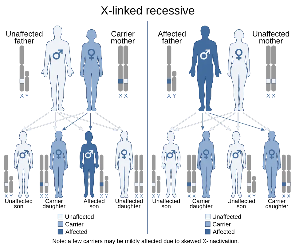
Carrier females may show a wide range of retinal findings because of random X-chromosome inactivation. Multimodal imaging can reveal mosaic RPE changes even in minimally symptomatic carriers.
Differential diagnosis
Choroideremia should be distinguished from other inherited retinal and chorioretinal disorders.
Retinitis pigmentosa
RP often shows bone-spicule pigmentation, retinal vessel attenuation, and variable inheritance. CHM is suggested by X-linked inheritance, choroidal atrophy, and exposed choroidal vessels.
Gyrate atrophy
Consider plasma ornithine testing and OAT analysis when sharply demarcated chorioretinal atrophy and autosomal recessive inheritance are suspected.
High myopia and myopic degeneration
Axial myopia, lacquer cracks, posterior staphyloma, and myopic choroidal neovascularization support myopic degeneration rather than CHM.
Bietti crystalline dystrophy and other retinal dystrophies
Crystalline deposits, corneal involvement, and autosomal recessive inheritance point toward Bietti crystalline dystrophy.
Management
No approved disease-modifying therapy currently cures CHM. Management focuses on monitoring, prevention of avoidable ocular stress, low-vision rehabilitation, and research readiness.
- • Regular ophthalmic review with OCT, FAF, and functional testing where available.
- • UV and glare protection, smoking cessation, and general vascular health.
- • Low-vision rehabilitation, orientation and mobility training, and assistive technology.
- • Genetic counselling for affected families and carriers.
Clinical research
Gene replacement therapy using AAV vectors carrying functional CHM has been the most advanced investigational approach. Trial outcomes have highlighted both the promise and the complexity of retinal gene therapy in CHM.
Other areas include outcome measure development, RNA-based strategies, genome editing, optogenetics, retinal prostheses, and cell-based approaches for advanced disease.
ClinicalTrials.gov SearchUseful resources
International resources
Questions about CHM?
Contact the patient community or refer patients for specialist ophthalmic genetic evaluation.